Your Heart and Heart Disease
Anatomy of the Heart
The heart is a muscle located in the middle of the chest and behind the breastbone (sternum) that is approximately the size of a fist. It is “powered” by an “electrical system” that signals the heart muscle to beat rhythmically approximately 72 times per minute.
If you were to slice it down the middle, you would find that it has three layers, the endocardium (the smooth inside lining of the heart); the myocardium (the muscle layer of the heart); and the epicardium (the outside lining of the heart). The pericardium is the tough, fluid-filled sac that surrounds the heart itself, this pseudo-fourth layer provides protection and minimizes the friction created by the heart beat.
The Heart is divided into four chambers: The Right Atrium (RA), the Right Ventricle(RV), the Left Atrium(LA) and the Left Ventricle (LV). The electraical signal for each heartbeat begins in the Right Atrium in an area called the sinus node (aka the heart’s natural pacemaker). Each chamber has a one-way valve at its exit that prevents blood from flowing backwards. When each chamber receives an electrical pulse, it contracts, and the valve at its exit opens pumping blood through it and when it is finished contracting the valve closes. As the lower chambers fill with blood, the electrical signal travels along special conduction tissues to the AV node, where it pauses for a few seconds, allowing the chambers to finish filling.
There are four valves in the heart including the Tricuspid Valve, which is at the exit of the Right Atrium, the Pulmonary Valve, which is at the exit of the Right Ventricle, the Mitral Valve, which is at the exit of the Left Atrium and the Aortic Valve, which is at the exit of the Left Ventricle.
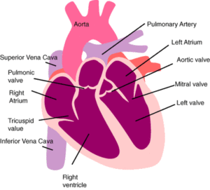
When the heart muscle contracts (or beats) it pumps blood out of the lower chambers of the heart. The heart contracts in two stages. In the first stage the Right and Left Atria contract at the same time, pumping blood to the Right and Left Ventricles. Then the Ventricles contract together (called systole) to propel blood out of the heart. After this second stage, the heart muscle relaxes (called diastole) before the next heartbeat. During this time, the muscle resets itself for contraction and blood fills the atria.
Functions of the Heart
The right side of the heart collects oxygen-poor blood from the body and pumps it to the lungs where it picks up oxygen and releases carbon dioxide while the left side collects oxygen rich blood from the lungs and pumps it to the body so that the cells throughout your body have the oxygen they need to function properly.
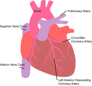
All blood enters the right side of the heart through two veins, the Superior Vena Cava(SVC), which collects blood from the upper half of the body and the Inferior Vena Cava (IVC), which collects blood from the lower half of the body.
When the heart is pumping, blood flows from the body to the Superior and Inferior Vena Cava then to the Right Atrium through the Tricuspid Valve. It flows to the Right Ventricle through the Pulmonary Valve through the Pulmonary Artery to the Lungs.
There, the blood picks up oxygen and drops off carbon dioxide in the lungs, and then flows from the lungs through the Pulmonary Veins to the Left Atrium through Mitral Valve to the Left Ventricle through the Aortic Valve to the Aorta through the two main coronary arteries — the Left Coronary Artery (which divides into two – the Left Anterior Descending Artery and the Circumflex Artery) and the Right Coronary Artery. From here blood flows the arterial system to the body.
The heart, just like any other organ, requires blood to supply it with oxygen and other nutrients so that it can do its work. The heart does not extract oxygen and other nutrients from the blood flowing inside it. The heart gets its blood from coronary arteries, located on the outside surface of the heart, that eventually carry blood within the heart muscle through a network of branches.
HEART FAILURE
Heart Failure does not mean your heart has stopped beating. It means that the heartbeat is not sufficient to supply an adequate volume of blood and oxygen to the brain and other parts of the body. When this occurs, a variety of compensatory changes will take place in an effort to pump an adequate amount of blood:
-the walls of the heart will stretch to increase volume capacity
-the walls of the heart will thicken to squeeze more forcefully
-the kidneys cause the body to retain sodium and water thereby increasing the amount of circulating blood
-hormones are released to make the heart squeeze more forcefully
Over time, these compensatory mechanisms will not be adequate to maintain sufficient circulation. The blood will not flow through the heart efficiently and as a result the heart becomes congested causing a backup of pressure in the circulatory system. This is known as Congestive Heart Failure (CHF) An associated build up of fluids occurs in the tissues, especially the lungs, causing peripheral swelling, fatigue and shortness of breath.
Two to three million Americans live with congestive heart failure. It is one of the most common reasons people 65 and older are admitted to the hospital. It can take years to develop.
Diagnosis
Symptomatology; blood tests; electrocardiography and echocardiography; x-rays, angiography.
Treatment
Treatment of chronic heart failure includes the use of multiple medications including: vasodilators (drugs that dilate blood vessels); ACE inhibitors (drugs that block vasoconstriction); inotropes (drugs that increase the heart’s ability to contract), and diuretics (drugs to reduce fluid). These medications are used alone and in combination.
Biventricular pacing
In a healthy heart, both upper chambers (atria) beat together as do both lower chambers (ventricles). Electrical impulses are delivered to the left ventricle in a highly organized pattern of contractions that efficiently pump blood out of the ventricle. In about one-third of patients with congestive heart failure (CHF), the electrical coordination is lost and the right and left ventricles do not beat together. This uncoordinated heart muscle function leads to inefficient ejection of blood from the ventricles and poses the risk of abnormal heart rhythms (arrhythmias). The biventricular pacemaker has leads implanted in the right atria, the right ventricle and the coronary sinus to sense and pace the left ventricle. This three-lead system allows the pacemaker to sense both ventricles and stimulate in a way that causes them to contract together. This resynchronization can help alleviate symptoms of CHF such as fatigue, shortness of breath and exercise intolerance thereby improving the patient’s overall quality of life.
Correction of underlying problem
Early diagnosis and corrective treatment of an underlying problem may minimize the risk of congestive heart failure. Medicines may be described to increase cardiac output and others to reduce volume overload.
Dietary regimen
Careful monitoring of the diet can help to keep symptoms of heart failure from flaring up. Heart failure patients eat a healthy diet that is low in salt and monitor their fluid intake.
Surgery
Corrective surgery is an option to treat underlying conditions to prevent them from leading to congestive heart failure. Surgery may also be done in some instances for the patient with congestive heart failure.
Surgical Treatments
Severe coronary artery disease (CAD) or valve disease may lead to CHF. Patients with CAD may benefit from angioplasty or bypass surgery. Patients with faulty heart valves can have valve replacement surgery. For severe CHF, a heart transplant may be needed.
Heart Failure Program – Multi-faceted Treatment:
The Congestive Heart Failure program at The University Hospital is designed to minimize the length of hospital stay for heart failure patients and to reduce admissions and readmissions. At the same time, the program also focuses on improving the patient’s ability to accomplish the routines of daily living as well as to reduce the number of medical complications associated with congestive heart failure.
The reduction of length of stay is accomplished through early and aggressive therapy for patients admitted with heart failure. Echocardiography is used in the Emergency department to make an immediate diagnosis so that therapy can start without delay. Admissions and re-admissions to the hospital are reduced because of a committed clinical staff who ensure that patients are seen frequently, adjustments to medications are made as needed, and will regularly re-enforce with patients the need for medical and dietary compliance.
The clinical staff help patients increase their ability to accomplish the routines of daily living by aggressively following up to be sure they take their medicines, and participate in a nutritional counseling and exercise rehab program. Patients who remain symptomatic despite receiving the best of conventional care are encouraged to participate in clinical trials.
The Program serves both hospitalized patients and as well as providing outpatient services by appointment at the NACC and Doctors Office Center (DOC).
Cardiac Disorders
Aortic Aneurysm
The aorta is the largest blood vessel in the body. All the oxygenated blood for the body is pumped out of the heart through this artery. It is then distributed to the body through a series of progressively smaller vessels. An aneurysm is a condition in which an area of an artery’s wall weakens and balloons out past its normal size. If the aneurysm occurs near the heart, in the aorta, it is called a thoracic (chest) aortic aneurysm; however, aneurysms can also develop in other parts of the body, such as the abdomen and the brain. Thoracic aortic aneurysms are relatively uncommon; most aneurysms-about 75 percent-occur in the abdomen. However, aortic aneurysms are prone to rupture once they reach a certain size. Fifty percent of patients who experience a rupture of a thoracic aortic aneurysm will die before reaching the hospital. Furthermore, surgical repair of a ruptured thoracic aneurysm carries a 25-50% mortality as opposed to a 5-8% mortality when such aneurysms are treated electively.
Diagnosis
Echocardiogram, computed tomography, or magnetic resonance imaging-and chest x-ray.
Treatment
Medication and lifestyle changes; surgery.
DISSECTING ANEURYSM
A major complication of aortic aneurysms is that they are prone to dissection or a tearing of the wall. An aortic dissection is an emergent condition in which the inner lining of the aorta tears allowing blood to pool between the inner and outer layers of the aorta. Potentially life threatening, this condition may require surgery to repair depending on the location of the aneurysm.
Diagnosis
Echocardiogram, computed tomography, or magnetic resonance imaging-and chest x-ray.
Treatment
Ascending aorta/Aortic arch – Emergent surgery
Descending aorta – blood pressure and pain control
ANGINA
Angina refers to the chest pain that is caused by an imbalance between the amount of oxygen the heart is receiving and the oxygen being consumed by the work that the heart is doing. Oxygen is delivered to the heart, through the coronary arteries, dissolved in the blood. The coronary arteries are small, approximately 2-4 millimeters in diameter. Over time, atherosclerotic plaque, an accumulation of calcium and cholesterol can build up in the blood vessels. As the plaque builds up in the coronary arteries, the passageway of the vessels becomes smaller thereby decreasing the blood supply feeding the heart. This causes the heart to receive less oxygen than it normally does. During physical activity or periods of stress, chemicals are released into the blood that cause the heart to beat faster. When the heart does more work it uses more oxygen and needs an increased blood supply. However, due to the atherosclerotic plaque, blood flow through the vessels is already at or close to capacity. Therefore the heart does not get the supply of blood and, as a result, oxygen that it needs. When deprived in this way, the heart complains by causing pain known as angina. This frequently occurs when extra demands are placed upon the heart, such as during exercise, stress, exposure to the cold, or even digestion of large meals.
Diagnosis
Symptoms; blood tests; stress tests; electrocardiography and echocardiography; angiography.
Treatment
Immediate: Cease physical activity, sit and rest Long term: Medications, angioplasty or surgery.
ARRHYTHMIAS/ABNORMAL HEART RHYTHMS
In order to function optimally, the heart must beat in a rhythmic pattern and at a controlled rate. However, there are conditions and disease states that cause the heart to beat irregularly or at an abnormal rate. It’s important for the heart to pump properly because that’s how your body gets the oxygen and food you need. A rate that is too slow may cause dizziness or cause a brief loss of consciousness (syncope.) A rate that is too fast may cause palpitations or shortness of breath. Electrophysiology is a specialized field that studies the relationship between the body’s function (in this case, the heart) and its electrical system. Examples of abnormal rhythms are:
Atrial Fibrillation (A fib)
A fib is a fast heart rhythm originating in the upper chambers of the heart in which an abnormal source of electrical stimulation causes the atrium to beat more rapidly and chaotically. Impulses are conducted to the ventricle in a random pattern and as a result, A fib is always an irregular rhythm. Sometimes people with atrial fibrillation can have a slow ventricular rate. Medicines used to control atrial fibrillation may result in slow rhythms, which are treated by pacemakers. Patients can live with A fib for a long time but it can lead to stroke or more serious heart rhythm disturbances.
Atrial flutter (A flutter)
A flutter is a fast heart rhythm originating in the upper chambers of the heart in which an abnormal source of electrical stimulation causes the atrium to beat more rapidly than normal. Not all impulses are conducted to the ventricle and as a result, the atria beat at a faster rate than the ventricles. A flutter may be regular or irregular and is less common than atrial fibrillation.
Ventricular tachycardia (v-tach)
V-tach is a fast heart rhythm originating in the ventricles (lower chambers) of the heart muscle. Although it is regular, the pumping action of the heart is inefficient and the rapid rate prevents the ventricles from filling adequately. As a result, circulation is severely reduced. This may cause a person to faint, drop their blood pressure and may lead to ventricular fibrillation. This is a life threatening rhythm that is most commonly associated with heart attacks.
Ventricular fibrillation (v-fib)
V-fib is an irregular heart rhythm, originating in the ventricles, in which there are very rapid, uncoordinated, fluttering contractions. This rapid chaotic movement does not allow the heart to contract properly and very little blood is pumped out to the body. This is a life threatening rhythm that is most commonly associated with heart attacks.
Heart blocks
This is a category of arrhythmias in which the electrical signals from the atria to the ventricles are delayed or not conducted at all. If no impulses are conducted the atria and ventricles will beat independently and at a different rate. As a result, less blood moves from the atria top the ventricle, the ventricles are not filled at the time of contraction and circulation is reduced.
Diagnosis
All heart rhythm disturbances are diagnosed by symptomatology and the use of electrocardiograms, Holter monitors, tilt table tests and electrophysiology studies
Treatment
Monitoring and lifestyle changes; medications (antiarrhythmic, heart rate control drugs and anticoagulants); pacemakers, implantable cardioverter-defibrillators (ICD); surgery.
HEART FAILURE
Heart Failure does not mean your heart has stopped beating. It means that the heartbeat is not sufficient to supply an adequate volume of blood and oxygen to the brain and other parts of the body. When this occurs, a variety of compensatory changes will take place in an effort to pump an adequate amount of blood: -the walls of the heart will stretch to increase volume capacity -the walls of the heart will thicken to squeeze more forcefully
-the kidneys cause the body to retain sodium and water thereby increasing the amount of circulating blood
-hormones are released to make the heart squeeze more forcefully
Over time, these compensatory mechanisms will not be adequate to maintain sufficient circulation. The blood will not flow through the heart efficiently and as a result the heart becomes congested causing a backup of pressure in the circulatory system. This is known as Congestive Heart Failure (CHF) An associated build up of fluids occurs in the tissues, especially the lungs, causing peripheral swelling, fatigue and shortness of breath.
Two to three million Americans live with congestive heart failure. It is one of the most common reasons people 65 and older are admitted to the hospital. It can take years to develop.
Diagnosis
Symptomatology; blood tests; electrocardiography and echocardiography; x-rays, angiography.
Treatment
Treatment of chronic heart failure includes the use of multiple medications including: vasodilators (drugs that dilate blood vessels); ACE inhibitors (drugs that block vasoconstriction); inotropes (drugs that increase the heart’s ability to contract), and diuretics (drugs to reduce fluid). These medications are used alone and in combination.
Biventricular pacing
In a healthy heart, both upper chambers (atria) beat together as do both lower chambers (ventricles). Electrical impulses are delivered to the left ventricle in a highly organized pattern of contractions that efficiently pump blood out of the ventricle. In about one-third of patients with congestive heart failure (CHF), the electrical coordination is lost and the right and left ventricles do not beat together. This uncoordinated heart muscle function leads to inefficient ejection of blood from the ventricles and poses the risk of abnormal heart rhythms (arrhythmias). The biventricular pacemaker has leads implanted in the right atria, the right ventricle and the coronary sinus to sense and pace the left ventricle. This three-lead system allows the pacemaker to sense both ventricles and stimulate in a way that causes them to contract together. This resynchronization can help alleviate symptoms of CHF such as fatigue, shortness of breath and exercise intolerance thereby improving the patient’s overall quality of life.
Correction of underlying problem
Early diagnosis and corrective treatment of an underlying problem may minimize the risk of congestive heart failure. Medicines may be described to increase cardiac output and others to reduce volume overload.
Dietary regimen
Careful monitoring of the diet can help to keep symptoms of heart failure from flaring up. Heart failure patients eat a healthy diet that is low in salt and monitor their fluid intake.
Surgery
Corrective surgery is an option to treat underlying conditions to prevent them from leading to congestive heart failure. Surgery may also be done in some instances for the patient with congestive heart failure.
Surgical Treatments
Severe coronary artery disease (CAD) or valve disease may lead to CHF. Patients with CAD may benefit from angioplasty or bypass surgery. Patients with faulty heart valves can have valve replacement surgery. For severe CHF, a heart transplant may be needed.
Heart Failure Program – Multi-faceted Treatment:
The Congestive Heart Failure program at The University Hospital is designed to minimize the length of hospital stay for heart failure patients and to reduce admissions and readmissions. At the same time, the program also focuses on improving the patient’s ability to accomplish the routines of daily living as well as to reduce the number of medical complications associated with congestive heart failure.
The reduction of length of stay is accomplished through early and aggressive therapy for patients admitted with heart failure. Echocardiography is used in the Emergency department to make an immediate diagnosis so that therapy can start without delay. Admissions and re-admissions to the hospital are reduced because of a committed clinical staff who ensure that patients are seen frequently, adjustments to medications are made as needed, and will regularly re-enforce with patients the need for medical and dietary compliance.
The clinical staff help patients increase their ability to accomplish the routines of daily living by aggressively following up to be sure they take their medicines, and participate in a nutritional counseling and exercise rehab program. Patients who remain symptomatic despite receiving the best of conventional care are encouraged to participate in clinical trials.
The Program serves both hospitalized patients and as well as providing outpatient services by appointment at the NACC and Doctors Office Center (DOC).
CORONARY ARTERY DISEASE
Coronary Artery Disease
Coronary artery disease (CAD) is a chronic disease in which atherosclerotic plaque (a combination of calcium and cholesterol) deposits along the walls of the coronary arteries leading to narrowing, hardening and stiffening of the arteries on the surface of the heart. CAD restricts blood flow through these vessels, preventing blood and oxygen from reaching areas of the heart. Due to this decrease in blood flow these areas become weak and do not function as well putting an increased burden on the rest of the heart. If not corrected, CAD can lead to angina, heart attack, heart failure or arrhythmias. CAD develops over time, is progressive and frequently has no symptoms till it is very advanced.
Diagnosis
Patient history and monitoring; blood tests; identification of risk factors such as elevated cholesterol levels, hypertension, smoking, diabetes and a lack of cardiovascular exercise; electrocardiography (EKG); stress testing (an EKG that is done while the patient is exercising in a controlled environment); echocardiography and/or cardiac catheterization.
Treatment
Medication and lifestyle changes that focus on the reduction/control of risk factors; balloon angioplasty; bypass surgery
HEART ATTACK
A heart attack, or a myocardial infarction (MI), occurs when the blood flow to a part of the heart is blocked. This happens because coronary arteries that supply the heart with blood slowly become clogged from a buildup of cells, calcium and cholesterol called plaque. This plaque can rupture (break apart). If it does, the blood flowing through these clogged arteries can form a clot. If this occurs, the clot cuts off the blood flow completely and the part of the heart muscle supplied by that artery begins to die. This can lead to arrhythmias, heart failure and septal defects. While you may only be in the hospital a few days, your recovery from a heart attack will be on going for the rest of your life. It is very important to take the steps necessary to prevent a second heart attack.
Diagnosis
EKG’s, blood tests, nuclear imaging, echocardiograms and cardiac catheterization.
Treatment
Medication and lifestyle changes; angioplasty; bypass surgery.
PULMONARY HYPERTENSION (PH)
Pulmonary hypertension is a rare blood vessel disorder of the lung in which the pressure in the pulmonary artery (the blood vessel that leads from the heart to the lungs) rises above normal levels and may become life threatening.
Diagnosis
Pulmonary hypertension is frequently misdiagnosed and has often progressed to late stage by the time it is accurately diagnosed. Pulmonary hypertension has been historically chronic and incurable with a poor survival rate. However, new treatments are available which have significantly improved prognosis.
Recent data indicate that the length of survival is continuing to improve, with some patients able to manage the disorder for 15 to 20 years or longer.
Treatment.
The treatment of primary pulmonary hypertension is complex. Patients benefit from referral to centers that specialize in the management of this uncommon problem.
Some orally administered drugs known as vasodilators, drugs such as calcium channel blockers, may alleviate pulmonary vasoconstriction and prolong life in about 20 percent of patients with primary pulmonary hypertension. In order to avoid significant adverse effects, patients are usually tested with potent, short-acting and titratable medication before a long-term therapy is selected.
A continuous infusion of a potent, short-acting vasodilator medication Epoprostenol (Flolan), or prostacyclin, is considered to be an important advance in the treatment of primary pulmonary hypertension.and most patients are able to learn how to prepare and infuse the drug. Patients may also be treated for chronic anticoagulation with warfarin (Coumadin)
SEPTAL DEFECTS
The heart is composed of four chambers, two atria (the upper chambers) and two ventricles (the lower chambers). The atria and ventricles are separated by the septum, a wall of muscle tissue that divides the heart right and left. A hole or holes may be present in this strip of muscle. These are known as septal defects.
Atrial Septal Defect (ASD)
In fetal circulation there is normally an opening between the two atria (the upper chambers of the heart) to allow blood to bypass the lungs. This opening usually closes about the time the baby is born. If this opening remains, it is known as an atrial septal defect. Blood will continue to flow from the left to the right atria – this is called a shunt.
ASD is present in 1 out of 25,000 people. When the person has no other congenital defect, symptoms may be absent, particularly in children. Symptoms usually have manifested by age 30. Individuals with ASD are at an increased risk for developing a number of complications including:
- infective endocarditis (a bacterial infection of the heart)
- right sided heart failure
- an abnormally fast heart rhythm known as atrial fibrillation
Diagnosis
Heart murmur on physical exam, EKG and echocardiography
Treatment
If the defect is small, no treatment may be necessary. If the patient becomes symptomatic, then closure is recommended, commonly by surgical means. Recently, a new non-surgical procedure has been developed to close the defect. This involves the introduction of a closure device known as an Amplatzer into the heart through the blood vessels. A tiny incision is made in the groin to introduce the catheters. They are then advanced into the heart where the closure devise is placed across the ASD and the defect is closed.
Ventricular Septal Defect (VSD)
Before birth, the right and left ventricles of its heart are not separate. As the fetus grows a muscular wall forms to separate these lower heart chambers. If the wall does not completely form, a hole remains. This is what is known as a ventricular septal defect, or a VSD.
It is estimated that up to 1% of babies are born with this condition. In the vast majority (80-90%) of babies born with this condition, the hole is small. They will have no symptoms, and the hole will close spontaneously as the muscular wall continues to grow after birth. If the hole is large, then too much blood will be pumped to the lungs, leading to congestive heart failure. These babies are often have symptoms related to the problem and may need medicine or surgery to close the hole.
In adults, interventricular septal defects are a rare, but serious complication of heart attacks. During a heart attack, the flow of blood is cut off to a section of the heart. If the blood flow is not reestablished within a few hours the effected tissue will die. If this effected tissue is in the septum, it is prone to rupture since the pressure in the left ventricle is much higher than the right. When there is a large opening between the ventricles, a large amount of oxygen-rich (red) blood from the heart’s left side is forced through the defect into the right side. Then it’s pumped back to the lungs, even though it’s already been refreshed with oxygen. This is inefficient, because already-oxygenated blood displaces blood that needs oxygen. This means the heart, which must pump more blood, may enlarge from the added work. High blood pressure may occur in the lungs’ blood vessels because more blood is there. Over time, this increased pulmonary hypertension may permanently damage the blood vessel walls.
Diagnosis
Presence of a murmur on physical exam, EKG, CXR, echocardiography.
Treatment
If the defect is small, it may never need intervention. However, if the opening is large enough to produce symptoms, then surgical repair is the only option. This requires open heart surgery, use of the heart lung bypass machine and closure of the defect, usually by insertion of a synthetic patch.
AORTIC STENOSIS
Aortic stenosis is one of the most common and serious valve disease problems. Aortic stenosis is a narrowing of the aortic valve opening. Because the narrowed your heart has to work harder to pump blood through the aorta and meet your body’s needs.
Many people with aortic stenosis don’t experience noticeable symptoms until the amount of restricted blood flow becomes greatly reduced. As the condition progresses and the valve gets narrower, you may experience chest pain, shortness of breath, dizziness or fainting. Over time, the this can weaken your heart and cause heart failure.
Our Heart Valve Disease Clinic at University Hospital brings together an expert interdisciplinary team including experienced interventional cardiologists and cardiothoracic surgeons to provide comprehensive care for patients with aortic valve stenosis. Our experts offer the full range of treatments, from minimally invasive procedures to open-heart surgery.