The Brain Tumor Program
During a FIGS procedure, fMRI is used to map the functional area of a patient’s brain. While the MRI is scanning, the patient is asked to perform a series of activities and movements, such as reading a list or tapping fingers. The areas of the brain that correlate to those movements “light up” on the scan and create an image. This information is sent to a surgical navigation computer located in the operating room. Neurosurgeons use a special pointer positioned on the patient’s head to guide incisions, skull openings, and brain tumors based on corresponding points of the MRI image. The added degree of precision in guidance and navigation provided by this technique maximizes tumor resection while minimizing the possibility of weakness, blindness, and speech loss.
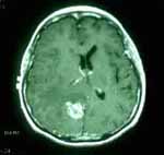
This image shoes a pre-operative MRI of a 48-year-old woman with a single metastic tumor from a small lung cancer.
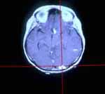
Using the Stealth Station, the fMRI shows the location of the visual cortex which is a distance away from the tumor.