

The Brain Tumor Program
Surgical resection of a brain tumor is a common and often effective treatment. But sometimes the tumor’s location near “eloquent” areas of the brain–those that control movement or speech–poses a dilemma. Removing the tumor may also result in taking away some of the healthy tissue surrounding it, causing a neurological deficit
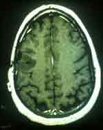
These images illustrate the case of a 34-year-old male with a recurrent benign brain tumor (a low grade glioma).This patient had previously had a biopsy and radiation treatment at another facility and was referred to the Brain Tumor Program for follow-up treatment. This image is a preoperative MRI.
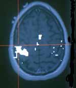
This image, taken using the Stealth Station technology shows the location of the motor cortex which is behind the tumor. Because of the precision of the brain mapping, the motor cortex and the tumor are distinguishable.
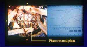
This image shows the stealth grid placed for brain mapping.
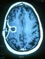
Finally, this MRI image is after the surgery and shows no tumor, and no damage to the motor cortex.